flow cytometry results for lymphoma
Ad BioAgilytix Global Labs Offers Assay Development Testing With The Latest Technologies. Morphology and immunohistochemistry on node tissue and bone marrow biopsies are frequently used in lymphoma diagnosis to characterize the stage and subtype of diseases.

Flow Cytometry Of Mantle Cell Lymphoma Shows Cd5 Positive B Cells Which Download Scientific Diagram
Flow cytometry provides important ancillary information in the diagnosis of lymphoma particularly when clinical or histology findings are limited equivocal or mimic malignancy.
. Ad Get A Treatment Plan That Best Targets Your Specific Cancer. Two-Color analysis primarily for surface markers is currently the standard method for flow cytometry measurements in routine diagnostic studies of leukemia and lymphoma. 5 segs 52 lymphocytes 32 monocytes 9 eosinophils.
Three samples that came from patients who had morphologic evidence of malignant disease on biopsy two Hodgkins disease and one large cell lymphoma had flow cytometry results that were interpreted as normal. Immunophenotyping Flow Cytometry for Hematolymphoid Neoplasia. Testing begins with decisions about which screen test panels to use for individual samples as they are received by the laboratory.
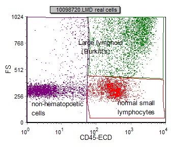
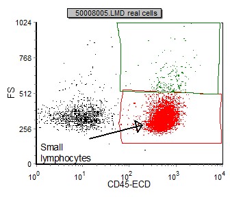
Flow cytometric immunophenotyping is useful in diagnosing lymphoma. These can be stratified as large and small lymphocytes CD45 positive. Flow cytometry has become an important tool in the diagnosis of mature lymphoid neoplasms and the determination of prognosis in selected cases.
Flow cytometry is generally used as follow up testing after a complete blood count CBC or white blood cells scan WBC. These cells were in the subsequent anlysis. 11 lymphs including hematogones Cytogenetics.
A broad range of immunophenotype patterns are interpreted for various type of leukaemia. With immunophenotyping your results will state whether any abnormal cells are present and what types of cells they are. Ad Lymphoma Is A Type Of Cancer That Affects The Lymph Nodes Of Your Body.
Flow cytometry analysis in brain biopsy is a feasible technique with 100 specificity to confirm the diagnosis of brain lymphoma in patients suspected for lymphoma on clinical grounds. Lineage identification can provide a confirmatory diagnosis or differential diagnosis prognosis and treatment options. Everyones Cancer Is Unique.
47XX8t922q34q112146XX19 t922 translocation in 1 of 200 cells analyzed. When using fresh tissue for flow cytometric immunophenotyping the predominant populations are lymphoid. Multicolor flow cytometry technology is a novel technique for the analysis of immunological markers to identify lymphoma on fresh tissue when immunohistochemical.
Your Support Should Be Too. However flow cytometry results usually make certain lymphoma entities extremely likely and others very unlikely. Leukemias and lymphomas are caused by an abnormal white blood cell that begins to divide uncontrollably making numerous copies of itself clones.
The typical patient is over the age of 50. The added clinical value is the speed by which flow cytometry can establish or confirm the diagnosis enabling a faster initiation of treatment while false positive cases were. This is especially true if initial testing showed an increased number of lymphocytes abnormal cell counts or the presence of immature blood cells.
Several recent studies that used either immunohistochemistry or molecular expression array profiling have demonstrated that specific patterns of the inflammatory milieu or antigen expression in HRS cells themselves correlate. The advantages of flow cytometry are based largely on its ability to analyse rapidly and simultaneously multiple cell properties in a quantitative manner. Not always strictly speaking not very often.
WBC1700uL Hb89 gdL Plt168000uL Differential count. The gating dot plot below identifies a predominant CD45 bright FS small used cells. It is postulated that SMZL may represent a large fraction of unclasssifiable CD5- chronic lymphocytic leukemias CLL.
Abstract of Lecture. We hypothesize that flow cytometry could also be successfully applied to prognostication of clinical outcome of Hodgkin lymphoma. Immunophenotyping is a type of flow cytometry used to diagnose leukemia or lymphoma.
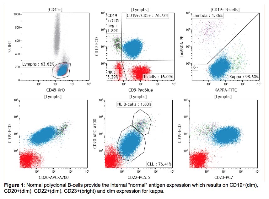
This test is usually done after abnormal results are seen on a complete blood count or WBC differential. Speak To Us Today To See How The Flow Cytometry Platform Can Move Your Molecules Forward. Flow cytometry is rapid and appears to be virtually diagnostic of non-Hodgkins lymphoma when a majority of cells are B cells with an abnormal kappalambda ratio.
This test generates a hematopathology report with a diagnosis and interpretation of findings. Flow cytometry immunophenotyping may be useful in helping to diagnose classify treat and determine prognosis of these blood cell cancers. Correlation of grade of lymphoma with flow cytometric CD19 forward scatter.
It is critical to recognise borderline cases in order to avoid misdiagnosing lymphoma and the current study estimates one-quarter of benign nodes exhibit outlier. After review of the clinical history and morphology a panel of markers is selected for each case by a board-certified hematopathologist. Therefore flow cytometry is an important integral part of lymphoma diagnosis even in cases where it cannot give a definitive diagnosis.
Grade 1 follicular lymphomas had a percentage of cells at or beyond the 500-channel mark ranging from 012 to 66 median 46 whereas grade 2 follicular lymphomas had a percentage ranging from 412 to 1255 median 7. The Lymphoma Warning Signs And The Many Faces Of It. It is used to detect abnormal hematolymphoid populations determine what cell surface markers they express and integrate immunophenotypic findings with morphologic and available clinical and.
The official flow cytometry laboratory report is most commonly an individual-lab. Flow cytometric leukemia and lymphoma analysis may aid in identifying the tumor lineage for diagnostic and prognostic purposes. Leukemia and lymphoma analysis by flow cytometry aids in identifying the tumor lineage which in most cases is identified as T cell B cell or myeloid.
MALT lymphomas account for approximately 75 percent nodal marginal zone accounts for less than 2 percent and splenic marginal zone lymphoma SMZL for less than 1 percent of NHL cases. Flow cytometry plays an important role in the diagnosis monitoring and treatment of haematological malignancies.

Flow Cytometric Immunophenotyping Performed On The Same Plasmablastic Download Scientific Diagram
Burkitt S Lymphoma Bl Flow Cytometry

Flow Cytometry Immunophenotypic Diagnosis Of B Cell Non Hodgkin Lymphomas On Fine Needle Aspirate Of Lymph Node Khanom Kh Tarafder S Sattar H Clin Cancer Investig J

Flow Cytometry Laboratory Duke Department Of Pathology

Selected Flow Cytometric Immunophenotyping Plots From Fine Needle Download Scientific Diagram

Summary Of Flow Cytometry Immunophenotypic Results For Anaplastic Large Download Table

Examples Of Cd200 Expression In Mantle Cell Lymphoma By Flow Cytometry Download Scientific Diagram

International Clinical Cytometry Society

Flow Cytometric Presentation Of A Large B Cell Lymphoma A Forward Download Scientific Diagram
Pathology Outlines Lymphoplasmacytic Lymphoma

Pb Flow Cytometric Analysis Download Table

Diffuse Large B Cell Lymphoma Dlbcl Flow Cytometry
Follicular Lymphoma Fl Flow Cytometry

Diffuse Large B Cell Lymphoma Dlbcl Flow Cytometry
International Clinical Cytometry Society
Follicular Lymphoma Fl Flow Cytometry

Flow Cytometry Immunophenotypic Diagnosis Of B Cell Non Hodgkin Lymphomas On Fine Needle Aspirate Of Lymph Node Khanom Kh Tarafder S Sattar H Clin Cancer Investig J

Immunophenotype By Flow Cytometry Of The Peripheral Blood Showing Download Scientific Diagram

International Clinical Cytometry Society